

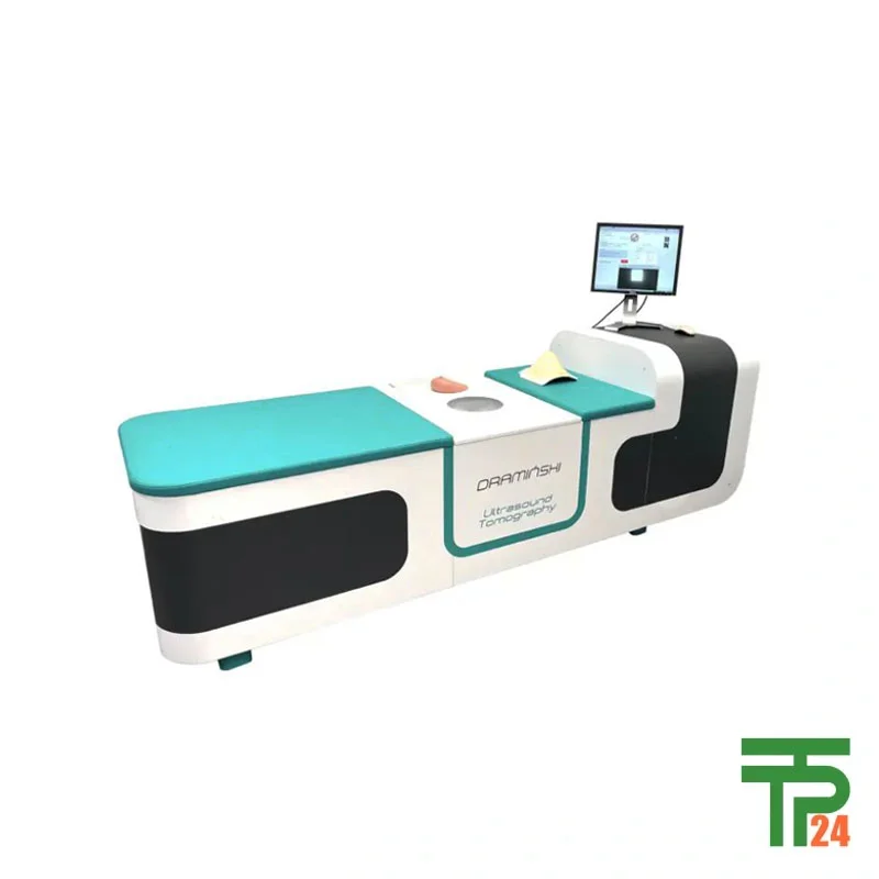
Draminski Ultrasound Tomography Breast Scanner
- Ultrasound frequency: ~2 MHz
- Transducers count: 1024 units
- Ring diameter: 260 mm
- Scan slice spacing: 1–2 mm
Did you like this product? Add to favorites now and follow the product.

(+86) 132-6243-8118 Start Live Chat
Description
Advanced Non-Invasive Breakthrough in Breast Diagnostics
The Draminski Ultrasound Tomography Breast Scanner represents a novel and groundbreaking approach in breast cancer diagnostics, combining non-invasive methodology with advanced hybrid technology. Designed and manufactured in Poland by DRAMIŃSKI S.A., this highly engineered medical imaging device employs ultrasound transmission tomography (UTT), enabling precise quantitative imaging of the internal breast tissue structure. By eliminating harmful radiation exposure and invasive procedures, it marks a revolutionary step in medical imaging technology. As a highly specialized diagnostic device, it uses its core omnidirectional imaging technology (FASCI) to blend multiple high-quality B-mode ultrasound images, thereby delivering comprehensive visual data for physicians to make informed decisions. This scanner aims to set a new international standard in 3D breast imaging by providing both clinical accuracy and patient-friendly procedures. Importantly, for healthcare professionals seeking authenticity and guaranteed quality, tpt24, the premier global online store, ensures worldwide delivery of the authentic DRAMINSKI Ultrasound Tomography Breast Scanner to any location with an exceptional quality guarantee.
Revolutionary Three-Dimensional Breast Imaging Technology
The device distinguishes itself with a full 3D volumetric imaging capability, utilizing advanced algorithms to reconstruct high-resolution, quantitative maps of ultrasound velocity (UTTc) and ultrasound attenuation (UTTα). These computed data sets present invaluable diagnostic indicators of breast tissue density and elasticity — both essential factors in differentiating between benign and malignant lesions. The inclusion of enhanced B-mode imaging (Full Angle Spatial Compound Imaging – FASCI) further increases image clarity by minimizing speckle noise and artifacts through sophisticated pixel averaging. Its efficiency stems from an intelligent scanning mechanism capable of acquiring 100–200 coronal slices for each breast depending on size, scanning one slice in approximately 6 seconds. This leads to complete scans within just a few minutes, a process that can be shortened by an estimated threefold with optimization. Rapid imaging makes it possible to evaluate results nearly in real-time, efficiently supporting clinical workflows.
Clinical Applications and Diagnostic Advantages Worldwide
This scanner shines in its core applications: early detection of breast cancer, differentiation between benign and malignant tumors, tumor localization, volumetric analysis, and monitoring of treatment response over time. It is applicable in hospitals, diagnostic centers, oncology departments, research facilities, and mobile diagnostic units. Due to its radiation-free nature and painless usage, it also becomes ideal for high-frequency screening in high-risk populations and for women with dense breast tissue, where traditional mammography proves less effective. Its ability to collect high-definition volumetric data enables clinicians to make more confident diagnoses while reducing patient anxiety from unnecessary biopsies. The tpt24 online store ensures healthcare institutions worldwide can access and procure this innovative system, complemented by full authenticity verification and international logistics support.
Safe, Comfortable, and Patient-Centered Examination Protocol
Patient comfort plays a vital role in the DRAMINSKI scanner’s appeal. The examination procedure requires the patient to lie on her stomach with the breast gently submerged into a tank of distilled water heated to approximately 25°C, which serves as the acoustic transmission medium. The system then performs automated coronal slice scanning every 1–2 mm, using a precisely engineered multi-element ultrasonic ring array with 1024 miniature piezoceramic transducers. These transducers, each with an active area of 0.5 × 18 mm, operate at a low resonant frequency of around 2 MHz to ensure deep penetration and accurate imaging. Since no breast compression is required, the discomfort usually associated with mammography is entirely eliminated. This non-invasive, contact-free method significantly enhances the patient experience, which in turn improves screening compliance rates in population-level breast health programs.
High-Performance Ultrasonic Ring Array Design
The ultrasonic ring array lies at the heart of the scanner’s superior imaging performance. Its circular configuration with a 260 mm diameter completely surrounds the breast, allowing uniform acquisition from multiple angles without repositioning. With each miniature transducer capable of working as both transmitter and receiver, the system can seamlessly switch between transmission mode and reflection mode, maximizing diagnostic output from a single scanning session. Transmission tomography produces attenuation and velocity maps of tissue properties, while reflection imaging via phased-array sub-apertures generates detailed structural maps comparable to conventional ultrasound findings. This dual-mode approach enhances diagnostic confidence, especially when identifying lesions with subtle acoustic differences that can be overlooked using single-mode imaging systems.
Advanced Imaging Modes for Enhanced Interpretation
The DRAMINSKI scanner supports Fusion Imaging: an integrated visualization where UTTc and UTTα maps are combined with FASCI structural ultrasound in a single image. Tumor-type classification is enhanced through color coding — fat tissue appears black, glandular blue, benign lesions yellow, and malignant lesions red. By incorporating patient age and breast density profiling, the device adjusts diagnostic thresholds dynamically for more personalized accuracy. This innovative image fusion, coupled with Multi-Planar Reconstruction (MPR) of coronal, sagittal, and transverse views, gives radiologists a complete anatomical and functional insight. In comparative studies, lesions confirmed via biopsy to be benign fibroadenomas were correctly mapped with high precision — a testament to the system’s diagnostic accuracy and potential to reduce unnecessary invasive procedures.
Competitive Edge over Conventional Modalities
Traditional breast imaging modalities, such as mammography and conventional B-mode ultrasound, are often limited by operator dependency, breast density effects, or ionizing radiation exposure. The DRAMINSKI Ultrasound Tomography Breast Scanner not only eliminates radiation risk but also offers quantitative tissue characterization capabilities missing in standard ultrasound. The FASCI methodology reduces image noise and improves overall resolution, while UTT provides a robust measure of tissue elasticity and structure unaffected by operator technique. Furthermore, its speed, comfort, repeatability, and volumetric storage of breast data make it ideal for longitudinal studies and comparative follow-ups. Institutions purchasing through tpt24 benefit from assured authenticity, comprehensive global shipping solutions, and a device engineered specifically to set a new benchmark in breast imaging diagnostics.
Future Potential and Global Implementation Strategy
Looking forward, the DRAMINSKI Ultrasound Tomography Breast Scanner holds promise for broader application in AI-assisted diagnostics, where automated lesion recognition and malignancy grading could be fully integrated into clinical routines. As the system evolves, scanning speed could be further reduced, increasing daily throughput in high-volume clinical centers. Additionally, its modular design supports planned software upgrades for improved imaging algorithms and enhanced user interface interaction. The device is poised to redefine global breast screening standards, especially in countries prioritizing non-invasive, radiation-free early cancer detection. With the international reach enabled by tpt24, healthcare providers from any country can adopt this cutting-edge technology with full confidence in its authenticity and backed by a global warranty — ensuring the highest standard of breast health diagnostics worldwide.
Specifications
| Specification | Value / Description |
|---|---|
| Imaging Method | Ultrasound Transmission Tomography (UTT) + Full Angle Spatial Compound Imaging (FASCI) |
| Transducer Count | 1024 miniature piezoceramic elements |
| Transducer Active Area | 0.5 × 18 mm |
| Operating Frequency | ~2 MHz |
| Ring Diameter | 260 mm |
| Slice Thickness Adjustment | 1 mm or 2 mm |
| Breast Immersion Medium | Distilled water, ~25°C |
| Scanning Modes | Transmission, Reflection, Fusion Imaging |
| Imaging Reconstruction | Multi-Planar Reconstruction (MPR) – Coronal, Sagittal, Transverse |
| Examination Duration | Several minutes per breast |
| Color Coding in Fusion Mode | Fat (black), Glandular (blue), Benign tumor (yellow), Malignant tumor (red) |
Data Sheet
Download Data Sheet Of Draminski Ultrasound Tomography Breast Scanner
Related products

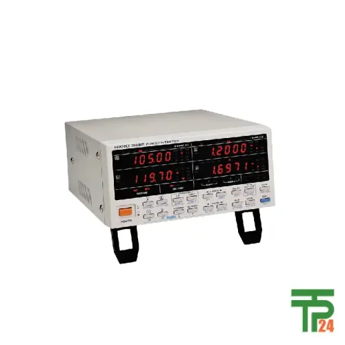
POWER HiTESTER 3332 HIOKI
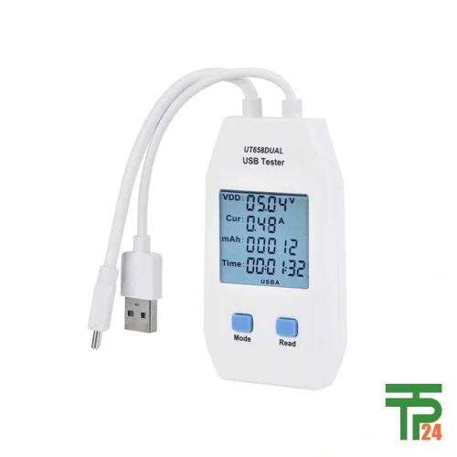
UNI-T UT658DUAL USB Testers
UNI-T UT658A USB Testers
Recently Viewed Products



Fair Pricing, Guarantee of Authenticity and Quality, Fast Delivery, and Provision of Technical Support to Customers.
Copyright 2025 © TPT24 Company. All right reserved. Powered by TPT24.
